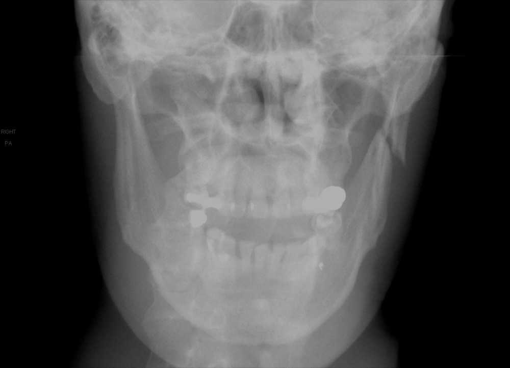
This condition may require surgical drainage. Unfortunately, when inflammation occurs, the mucous membrane swells and closes the aperture, this blocks normal sinus drainage.

One of the largest problems with sinuses is inflammation, which can be caused by numerous problems including infection and structural abnormalities, and in itself can cause pain and increased infections. With the exception of the lacrimal and palatine sinuses which are diverticula of the maxillary sinus, each sinus has a direct opening into the nasal cavity. Therefore, the lining of each sinus comprises of respiratory epithelium. The paranasal sinuses develop via evagination into the spongy bone between the external and internal plates of the cranial and facial bones.

This chapter will therefore be useful as a normal reference guide for clinical applications. The anatomical features (including head bones, muscles, and soft tissues) have been compared using both dissected heads and skulls and computed tomography images. Topographic descriptions and the relationships between the various air cavities and paranasal sinuses have been visualized using computed tomography and cadaver sections images. The purpose of this chapter is to present an anatomical reference guide of the paranasal sinuses in domestic animals, including large and small ruminants (cattle, buffalo, sheep, and goats), camels, canines (dog) and equines (horse and donkey), appropriate for use by anatomists, radiologists, clinicians, and veterinary students. Thus, each sinus is lined by respiratory epithelium and has direct or indirect communication to the nasal cavity. Paranasal sinuses are paired cavities within the skull, which develop by evagination into the spongy bone between the external and internal plates of the cranial and facial bones.


 0 kommentar(er)
